
Knowledge base
Hidden root canals: why a tooth can keep causing problems after root canal treatment
Why a root canal sometimes goes unnoticed, and how targeted diagnostics, the surgical microscope and, where needed, CBCT can help with persistent symptoms after root canal treatment.

Author:
Danilo Di Vico
Dentistry, surgery and digital implant dentistry at 3D Tandarts Schiedam.
Last updated: 30 September 2026
Reading time ~ 15 minutes
Do you still have pain in a tooth after root canal treatment? Or do symptoms return months or years later, such as tenderness when biting, swelling or an infection at the root tip?
That does not automatically mean the earlier root canal treatment was done incorrectly. There can be several causes. One of them is a root canal that was not found or treated during the earlier procedure.
The anatomy inside a tooth can be considerably more complex than a standard X-ray shows. Some teeth have extra canals, canals can split and rejoin, and small canal entrances can be hidden under dentine or hard to see because of calcification.
In this article we explain why root canals are sometimes missed, which teeth are more often affected, what this can mean for the outcome of a root canal treatment or a root canal retreatment, and how microscopy and targeted 3D diagnostics (CBCT) can help in complex cases.
Short answer
Persistent or recurring symptoms after root canal treatment can have several causes. A missed or untreated root canal is one of them.
Targeted clinical examination and X-ray diagnostics are therefore the first step. In complex anatomy, treatment under a surgical microscope can help to reveal small anatomical details. When conventional two-dimensional X-rays do not adequately answer a relevant diagnostic question, additional CBCT imaging may be considered in selected cases.
Contents
In this article
- When might a hidden canal be suspected?
- Which teeth have complex canal anatomy?
- Why does a missed canal matter?
- Microscope and CBCT
- Can a missed canal be treated afterwards?
- Frequently asked questions
When might a hidden root canal be suspected?
A missed canal is only one of several possible explanations for symptoms after an earlier root canal treatment.
Reasons for further investigation include:
- persistent or recurring pain in a previously treated tooth;
- tenderness or pain when biting;
- recurrent swelling or a fistula (gum boil);
- an infection at the root tip that does not heal or becomes visible again;
- symptoms that appear months or years after the original treatment;
- doubts about the anatomy or quality of the earlier treatment.
It is important to understand that these signs do not prove that a canal was missed.
A persistent infection in the treated canal system, coronal leakage, a root fracture, a restorative problem or a combined endodontic-periodontal condition can also cause similar symptoms.
That is why any retreatment starts not with treatment, but with diagnostics.
Still having symptoms after root canal treatment?
Is a tooth still tender, painful or inflamed after earlier root canal treatment? We can investigate the possible cause and whether retreatment makes sense.
Why do hidden root canals exist?
A root canal system is not a collection of perfectly straight tubes.
The internal anatomy forms during tooth development and can vary greatly. Canals can split, rejoin, be linked to one another by isthmuses, or have an irregular shape.
Some root canals also have a very small entrance. Others lie beneath a ledge of dentine or become harder and harder to see as secondary or tertiary dentine forms.
This means that two teeth that look almost identical from the outside can have different internal root canal anatomy.
Which teeth more often have complex canal anatomy?
Although anatomical variation can occur in almost any tooth, some groups of teeth are particularly known for it.
Upper first molars and the MB2 canal
A well-known example is the upper first molar.
Besides the first mesiobuccal canal, a second mesiobuccal canal can be present in the same root: the so-called MB2 canal.
A systematic review and meta-analysis by Martins et al. found a pooled MB2 prevalence of 69.6% (95% confidence interval 64.5–74.8%) in upper first molars. In upper second molars the pooled prevalence was lower, at 39.0%.
That does not mean a separate MB2 canal is clinically accessible or treatable in every upper first molar. It mainly shows how variable the anatomy of this tooth can be.
The MB2 canal can be small and its entrance can be hidden under dentine. That is why, when upper molars are treated, this anatomical variation is deliberately looked for.
Lower molars
Lower molars can also show considerable variation.
Besides the usual mesial and distal canal systems, extra canals can be present. The canals can also split, merge or be connected by isthmuses.
Lower incisors and premolars
A tooth that appears on a two-dimensional X-ray to have one root and seemingly one canal does not necessarily consist of a single canal inside.
Especially in certain lower incisors and premolars, two canals can be present that run close together or split further down the root.
C-shaped canal systems
In some molars, particularly certain lower second molars, the canal anatomy can be C-shaped.
Instead of several clearly separate round canals, there is a complex, ribbon-shaped canal system. This affects both locating the canals and cleaning and disinfecting the entire canal system.
Why is a missed root canal clinically relevant?
The aim of a root canal treatment is to clean and disinfect the infected root canal system as completely as possible and then seal it.
If a canal is not found and remains untreated, infected tissue and bacteria can remain in it. This can contribute to persistent or later recurring apical inflammation.
This link has also been found in scientific research.
Karabucak et al. examined root-filled teeth with CBCT and found that teeth with a missed canal were 4.38 times more often associated with a periapical lesion than teeth in which no missed canal was identified.
A recent systematic review and meta-analysis by León-López et al. from 2025 likewise found a significant association between missed canals detected on CBCT and post-treatment apical periodontitis. The authors also point to considerable heterogeneity between the included studies and a low certainty of the available evidence. The association is therefore clinically relevant, but it must not be interpreted as if every missed canal automatically causes symptoms or a lesion.
A missed canal is also not the only possible cause of persistent symptoms after root canal treatment.
This distinction matters: the diagnosis must be established before deciding whether retreatment is genuinely worthwhile.
Why are some root canals not found?
There are several possible reasons.
Limited visibility
Canal entrances can be very small and lie deep in the pulp chamber. In addition, overhangs of dentine can hide anatomical structures from direct view. A surgical microscope can largely overcome this problem; we explain how it works further on.
Anatomical variation
A clinician must not only know what the most common anatomy looks like, but also actively allow for possible variants.
An extra canal is not necessarily located where an opening would be expected based on standard anatomy.
Calcification
Age, restorations, trauma or earlier irritation can lead to extra dentine being formed. As a result, access to a canal that was originally present can become narrower and harder to recognise.
Limitations of two-dimensional X-rays
A conventional X-ray projects a three-dimensional structure onto a two-dimensional image.
Anatomical structures that lie one behind the other in the direction of the X-ray beam can therefore be projected over one another. An extra canal can thus be present without being separately recognisable on a standard image.
How do we look for hidden root canals?
Treatment under the surgical microscope
A surgical microscope combines strong magnification with focused illumination of the treatment area.
This makes it easier to assess small anatomical features of the floor of the pulp chamber, such as subtle colour differences, developmental grooves and small canal entrances.
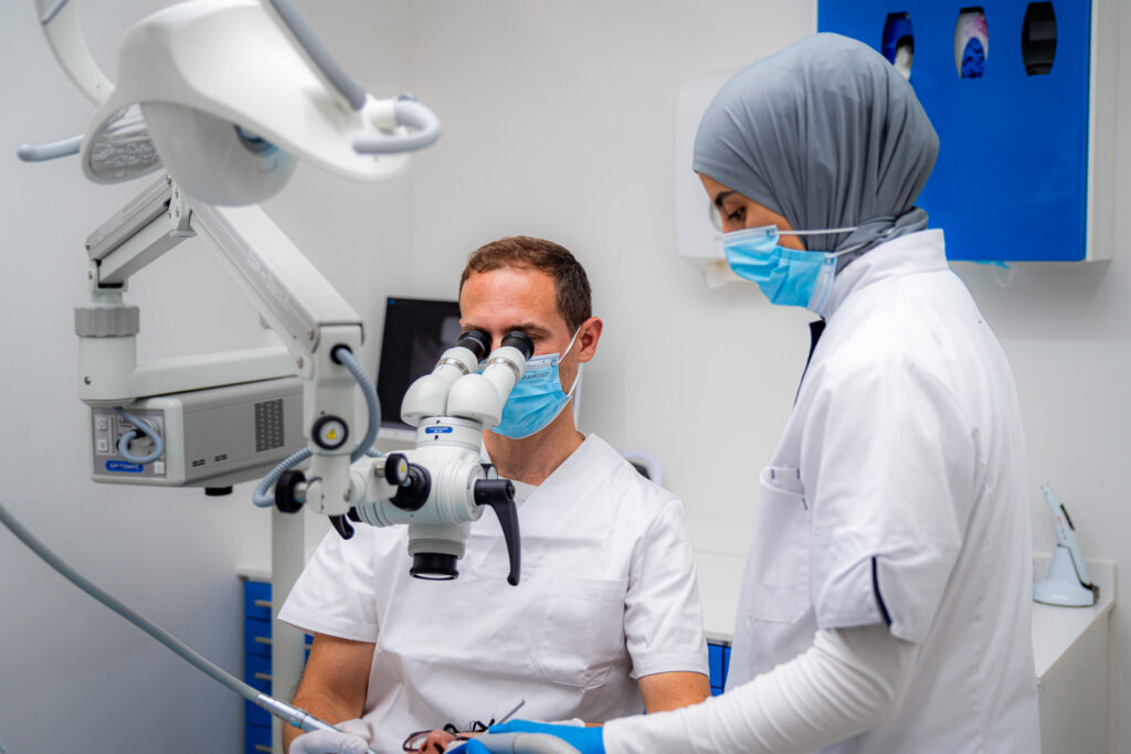
At 3D Tandarts we carry out root canal treatments and retreatments under a surgical microscope.
The microscope is not a substitute for anatomical knowledge or a systematic treatment strategy. It is an instrument that allows the clinician to see the anatomy better and investigate it more precisely.
Systematic examination of the pulp chamber
Magnification alone is not enough.
The floor of the pulp chamber is assessed systematically for anatomical clues to additional canals. The position of canal entrances already found, developmental grooves and colour differences in the dentine can all give direction.
In teeth known for complex anatomy, we actively allow for possible extra canals.
When can a CBCT scan help?
A CBCT scan allows a three-dimensional assessment and can provide additional diagnostic information in selected complex endodontic situations.
However, that does not mean a CBCT is automatically needed for every root canal treatment or retreatment.
The AAE and AAOMR do not recommend CBCT as a routine screening examination when conventional diagnostics provide enough information. CBCT is mainly relevant when a specific clinical question cannot be adequately answered with clinical examination and conventional two-dimensional X-rays.
This may be the case, for example, with:
- suspected complex or unusual root canal anatomy;
- persistent symptoms whose cause remains unclear with conventional diagnostics;
- suspicion of a missed canal;
- certain suspected resorptions or root fractures;
- planning of a complex endodontic retreatment or endodontic surgery.
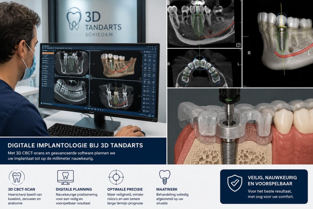
At 3D Tandarts we have a CBCT scanner on site. This means additional three-dimensional diagnostics, when there is genuinely an indication for them, can be part of the same diagnostic workflow.
A CBCT does not replace clinical inspection under the microscope. The two techniques provide different information and can complement each other in selected complex situations.
Is the cause of your symptoms still unclear?
With complex endodontic symptoms, we first assess what is going on. If necessary, microscopic examination and additional 3D diagnostics can be part of that assessment.
Can a missed root canal still be treated?
In many cases a missed canal can still be found and treated during a root canal retreatment.
A retreatment, however, is not simply a repeat of the original treatment.
First we need to establish why the earlier treatment did not achieve a sufficient result. We then assess whether the existing restoration is accessible, how the root canal anatomy runs, whether there are technical complications and what the prognosis of the tooth as a whole is.
When retreatment is worthwhile and technically possible, the existing canal fillings are removed where necessary, access to the canal system is regained and untreated anatomy is actively sought.
The aim remains the same: to preserve the natural tooth when that is justified and predictably possible.
Does pain after root canal treatment mean the treatment has failed?
No.
Short-term tenderness after a root canal treatment can occur and does not in itself mean the treatment has failed. Symptoms that appear later also have several possible causes.
So the question is not only:
“Does the tooth still hurt?”
but above all:
“Why does this tooth hurt?”
Only once the cause is sufficiently clear can it be decided whether waiting, restorative treatment, endodontic retreatment, endodontic surgery or another approach is most appropriate.
Frequently asked questions
Frequently asked questions
Does everyone have an MB2 canal?
No. Anatomy differs from person to person and from tooth to tooth.
In upper first molars an MB2 canal is indeed common. A systematic review and meta-analysis reported a pooled prevalence of 69.6%, but its presence does not automatically mean the canal runs the same way or is clinically accessible in every individual tooth.
Can a hidden root canal be missed on an ordinary X-ray?
Yes. A conventional X-ray is two-dimensional. Structures can be projected over one another, so complex three-dimensional anatomy is not always separately visible.
Does persistent pain always mean a canal was missed?
No. A missed canal is only one possible explanation.
A persistent infection, leakage, a restorative problem, a root fracture or another condition can also cause symptoms. That is why a targeted diagnosis is necessary before retreatment is recommended.
Can a missed canal still be treated?
In many cases, yes.
Whether a root canal retreatment is possible and sensible depends on factors including the anatomy, the existing restoration, any technical complications and the prognosis of the tooth.
Does a hidden root canal always cause symptoms?
No, not always.
An anatomically present but untreated canal does not necessarily lead to symptoms straight away. When a persistent infection is present, however, apical inflammation can develop or persist. Sometimes this is discovered because symptoms appear; sometimes an abnormality is seen on X-ray diagnostics.
Is a CBCT always needed for retreatment?
No, not always.
CBCT is not taken routinely for every root canal treatment or retreatment. It is considered when the additional three-dimensional information is expected to be relevant to a specific diagnostic or therapeutic question.
Is every root canal treatment at 3D Tandarts performed under a microscope?
At 3D Tandarts, root canal treatments and retreatments are carried out under the surgical microscope. The combination of magnification and focused illumination helps in assessing small anatomical details inside the tooth.
Symptoms after an earlier root canal treatment?
When a previously treated tooth causes problems again, it is important to establish why first.
At 3D Tandarts Schiedam we can reassess a previously treated tooth. We combine clinical examination and digital X-ray diagnostics with examination and treatment under the surgical microscope. When additional three-dimensional diagnostics are indicated, we have a CBCT scanner on site.
Based on the findings, we discuss whether a root canal retreatment is possible and worthwhile, or whether another treatment suits the situation better.
Still in pain from a tooth after root canal treatment?
Have the source of the symptoms investigated first. We assess the tooth and then discuss which treatment, if any, is most appropriate.
Medical information and disclaimer
The information on this page is intended as general dental information and does not replace an individual examination, diagnosis or treatment advice.
Persistent or recurring symptoms after root canal treatment can have several causes. The presence of symptoms does not automatically mean that a root canal was missed or that retreatment is necessary.
Which diagnostics and treatment are appropriate is determined for each patient on the basis of their symptoms, clinical findings, available imaging, restorative situation and the prognosis of the tooth concerned.
Sources and further information
The content of this article is based on clinical endodontic principles and has been checked against scientific literature and professional guidelines.
Scientific sources
Martins JNR, Alkhawas MAM, Altaki Z, et al. Second mesiobuccal root canal in maxillary molars — A systematic review and meta-analysis of prevalence studies using cone beam computed tomography. Archives of Oral Biology. 2020;113:104589.
For upper first molars, the meta-analysis reported a pooled MB2 canal prevalence of 69.6% (95% CI 64.5–74.8%) and for upper second molars 39.0% (95% CI 31.1–46.9%).
View publication on PubMed
Karabucak B, Bunes A, Chehoud C, Kohli MR, Setzer F. Prevalence of Apical Periodontitis in Endodontically Treated Premolars and Molars with Untreated Canal: A Cone-beam Computed Tomography Study. Journal of Endodontics. 2016;42(4):538–541.
In this CBCT study, teeth with a missed canal were 4.38 times more often associated with a periapical lesion.
View publication on PubMed
León-López M, et al. Association Between the Presence of Missed Canals, Detected Using CBCT, and Post-Treatment Apical Periodontitis in Root-Filled Teeth: A Systematic Review and Meta-Analysis. Journal of Clinical Medicine. 2025;14(16):5781.
The systematic review and meta-analysis found a significant association between missed canals and post-treatment apical periodontitis. The authors also reported high heterogeneity between the studies and a low certainty of evidence.
View publication on PubMed
CBCT guidelines
American Association of Endodontists (AAE) & American Academy of Oral and Maxillofacial Radiology (AAOMR). Joint Position Statement: Use of Cone Beam Computed Tomography in Endodontics.
CBCT is not a routine screening examination for every endodontic patient. The indication is determined by the clinical question and by whether conventional diagnostics provide enough information.
AAE/AAOMR position statement
Patient information
For general Dutch patient information about root canal treatments, you can also consult the information from the KNMT.
KNMT – Root canal treatment
Last updated: 30 September 2026

BIG registration: 29919697202
Knowledge base
Latest articles

Surgical microscope for root canal treatment in Schiedam
What does a surgical microscope do in root canal treatment? See how precision and CBCT help save your tooth. Book at 3D Tandarts in Schiedam.
Implant consultation in Schiedam: find out which treatment suits you
Is a dental implant right for you? Find out what an implant consultation in Schiedam involves: examination, advice and a treatment plan. Book today.
Night guard in Schiedam for teeth grinding and jaw clenching
Struggling with teeth grinding or jaw clenching? Learn when a custom night guard makes sense and why diagnosis matters. Book an assessment in Schiedam.
Questions about your situation?
During a consultation we look at your situation and discuss the options.